This article was published in Scientific American’s former blog network and reflects the views of the author, not necessarily those of Scientific American
Late last month the prominent neuroscientist Marian C. Diamond passed away at age 90. She is known for her study of how certain types of experiences can induce structural and chemical changes in the brain.
Like many of the most important scientists of the last century, Diamond’s publications included an article for Scientific American. Co-authored with her colleagues Mark R. Rosenzweig and Edward L. Bennett, the story “Brain Changes in Response to Experience” was featured on the cover of the February 1972 issue.

Credit: Bunji Tagawa
On supporting science journalism
If you're enjoying this article, consider supporting our award-winning journalism by subscribing. By purchasing a subscription you are helping to ensure the future of impactful stories about the discoveries and ideas shaping our world today.
In their experiments, Diamond and company placed rats in different sorts of environments ranging from solitary and boring to crowded and highly stimulating. Then they looked at how the rats’ brains came to differ from one experimental group to another. Their findings helped to lay the foundation for what we now know as plasticity in the human brain. As in many Scientific American articles published in the 1970s, a series of drawings by scientific illustrator Bunji Tagawa helped to tell the story. The images below provide an overview of the experimental setup and key results. You can also find the full article in our archive.
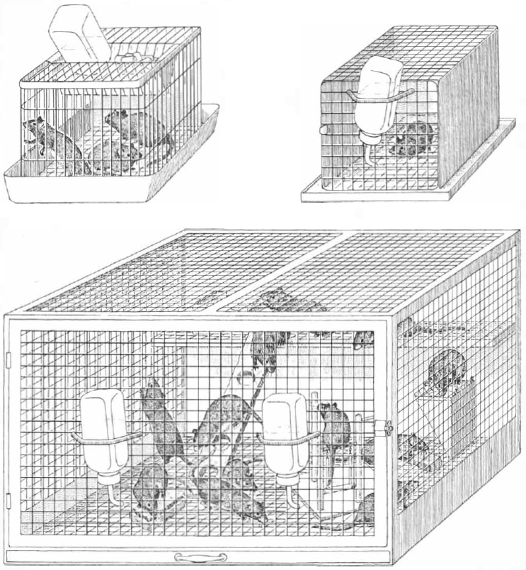
Three laboratory environments that produce differences in brain anatomy of littermate rats are depicted. In the standard laboratory colony there are usually three rats in a cage (upper left). In the impoverished environment (upper right) a rat is kept alone in a cage. In the enriched environment 12 rats live together in a large cage furnished with playthings that are changed daily. Food and water are freely available in all three environments. The rats typically remain in the same environment for 30 days or more.
Credit: Bunji Tagawa
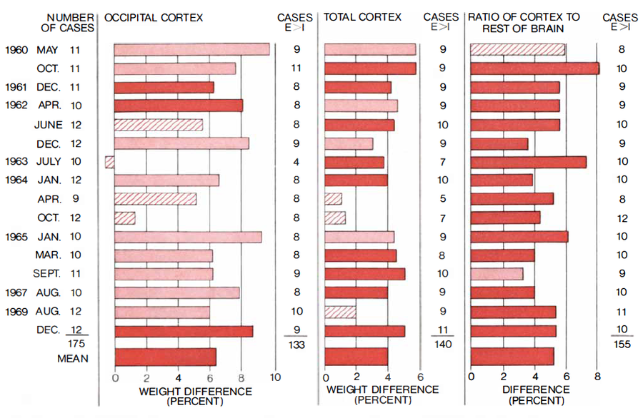
Brain weight differences between rats from enriched environments and their littermates from impoverished environments were replicated in 16 successive experiments between 1960 and 1969 involving an 80-day period and the same strain of rat. For the occipital cortex, weight differences in three of the replications were significant at the probability level of .01 or better (dark colored bars), nine were significant at the .05 level (light colored bars) and four were not significant (hatched bars). The ratio of the weight of the cortex to the rest of the brain proved to be the most reliable measure, with 14 of the 16 replications significant at the .01 level.
Credit: Bunji Tagawa
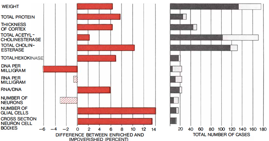
Occipital cortex of rats kept in enriched or impoverished environments from 25 to 105 days showed the effects of the different experiences. The occipital cortex of rats from the enriched environment, compared with that of rats from the impoverished one, was 6.4 percent heavier. This was significant at the .01 level or better, as were most other measures (dark colored bars). Only two measures were not significant (hatched bars). The dark gray bars on the right show how the number of cases in which the rat from the enriched environment exceeded its littermate from the impoverished environment in each of the measures that are listed.
Credit: Bunji Tagawa
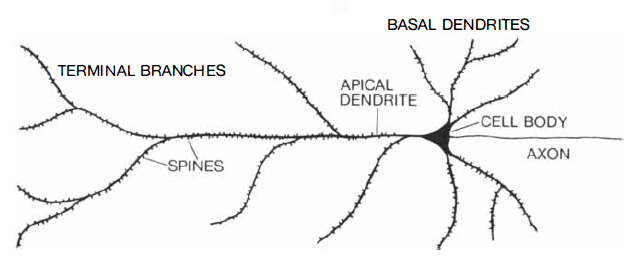
Dendritic spines, tiny “thorns,” or projections from the dendrites of a nerve cell, serve as receivers in many of the synaptic contacts between neurons. The drawing is of a type of cortical neuron known as the pyramidal cell. Rats from an enriched environment have more spines on these cells than their littermates from an impoverished environment.
Credit: Bunji Tagawa
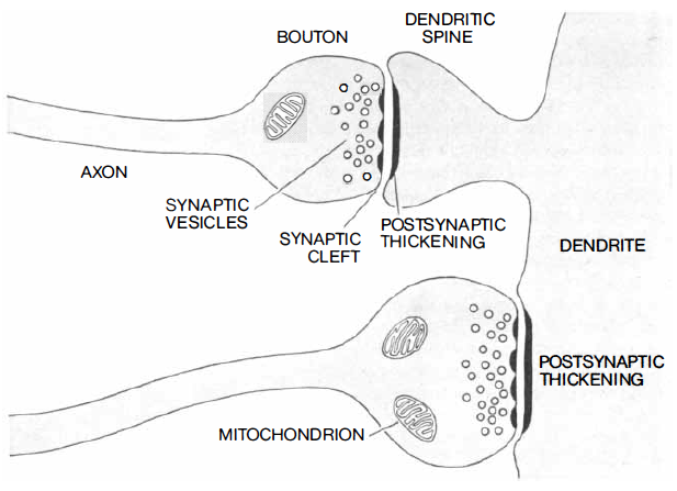
Synaptic junctions between nerve cells can be between axon and dendritic spine or between axon and the dendrite itself. The vesicles contain a chemical transmitter that is released when an electrical signal from the axon reaches the end bouton. The transmitter moves across the synaptic cleft and stimulates the postsynaptic receptor sites in the dendrite. The size of the postsynaptic membrane is thought to be an indicator of synaptic activity.
Credit: Bunji Tagawa
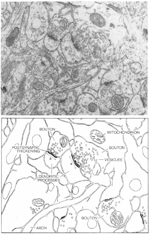
Brain section from the occipital cortex of a rat is enlarged 37,000 times in this electron micrograph by Kjeld Møllgaard. The map identifies some of the components. Measurement of the postsynaptic thickening is shown in the map of the section by the arrow (a). The number of synaptic junctions was also counted. It was found that rats reared in an enriched environment had junctions approximately 50 percent larger than littermates from an impoverished environment, and the latter had more junctions, although smaller ones, per unit area.
Credit: Kjeld Møllgaard (micrograph); Bunji Tagawa (map)