This article was published in Scientific American’s former blog network and reflects the views of the author, not necessarily those of Scientific American
The biology and behaviour of large theropod dinosaurs remains one of the most discussed and popular subjects within the entirety of vertebrate palaeontology, and continuing discussions demonstrate that the life appearance and external soft tissue anatomy of non-bird dinosaurs remain of equal popularity too. Today sees the publication of a new study – led by Chris Barker and involving myself and a team of co-authors – that has some bearing on both of these issues. The work revolves around the Early Cretaceous allosauroid Neovenator salerii from the Wessex Formation of the Isle of Wight, and specifically its facial anatomy.
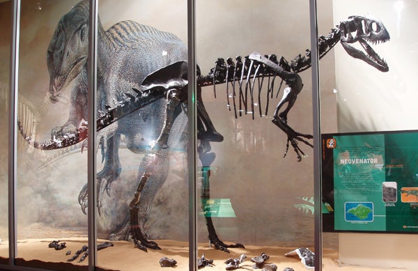
The fantastic holotype skeleton (NHMUK R10001/MIWG 6348) on display at Dinosaur Isle Museum, Sandown, Isle of Wight. Photo by Steve Brusatte, from Naish (2011). Credit: Naish 2011
Unusually for British theropods, Neovenator includes an extremely well preserved, three-dimensional partial skull (Brusatte et al. 2008), and it was the well preserved nature of this material (combined with its proximity and availability) that inspired us to study it anew. Working at the University of Southampton’s µVIS X-ray Imaging Centre (‘µVIS’ is said Mu-Vis, if you’re wondering), Chris Barker, myself and colleagues opted to CT-scan the specimen in an effort to discover something new about the vasculature, detailed bone anatomy and pneumaticity of this dinosaur’s face. Our new study – Barker et al. (2017) – appears today in Scientific Reports, and I’m pleased to report that it’s open access.
On supporting science journalism
If you're enjoying this article, consider supporting our award-winning journalism by subscribing. By purchasing a subscription you are helping to ensure the future of impactful stories about the discoveries and ideas shaping our world today.
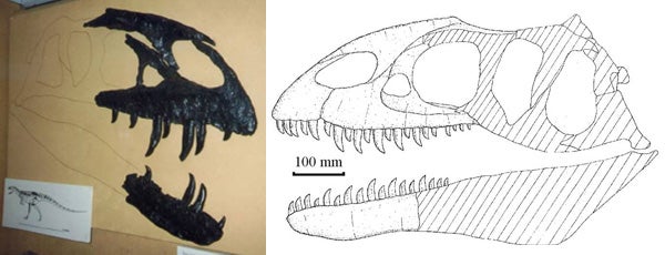
At left: the Neovenator skull as it used to be on display at the old Museum of Isle of Wight Geology during the late 1990s. At right: Steve Hutt's reconstruction of the skull, from Steve's MPhil thesis and also used in Naish et al. (2001). Credit: Darren Naish, Naish et al. 2001
By now I’ve been involved in several CT-scanning projects. They’re often a bit of a gamble; sometimes the results are poor and sometimes they’re terrible. On this occasion, I’m pleased to report that they were excellent. We were immediately struck by the extensive, complex, anastomosing system of internal channels and branches present deep inside the premaxillae and maxillae, snaking and curving around the tooth roots. After eliminating the possibility that these structures might be something crazy like invading plant roots or an artefact of the scanning process, we confirmed that they’re exactly what they look like: evidence for a well developed neurovascular system that deeply invades the bones and is connected to a series of external openings, termed foramina (Barker et al. 2017).
.jpg?w=600)
The left premaxilla and maxilla of Neovenator, showing the internal structure. The dotted lines show where vertical sections were taken (they're illustrated in the paper). Credit: Barker et al. 2017
The connection of the channels with external foramina known in living reptiles to be the passageways for nerves, their separation from the pneumatic system, their overall morphology and size, and their position relative to the facial features of living reptiles all show that these structures correspond to parts of the nervous system – we think that they’re branches of the trigeminal nerve, in particular the ophthalmic and maxillary divisions (Barker et al. 2017). We worked out how large these nerves are (in volume) and what percentage of the bone they actually occupy. They appear to have been pretty big in comparison with the facial nerves of other animals. We do think that various of the facial arteries and veins occupied similar positions to these nerve branches, though it’s hard to say precise things about them.
These big, invasive nerves provide – we think – evidence that Neovenator had a ‘sensitive’ face; that it was well able to receive a large amount of sensory information – pertaining to touch and perhaps temperature, pressure and other stimuli as well – via the sides and front of its face (Barker et al. 2017). It’s very tempting to think that its sensory abilities were similar to those of crocodylians where an enlarged trigeminal nerve – combined with structures termed dermal pressure receptions and integumentary sense organs – give these animals tactile and sensory discrimination perhaps “greater than primate fingertips” (Leitch & Catania 2012), as well as abilities to collect thermal and chemosensory information (Di-Poï & Milinkovitch 2013).
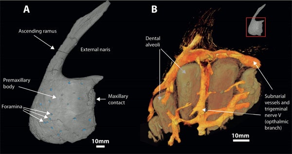
A. Neovenator left premaxilla in lateral view, foramina marked in blue. B. The premaxillary body with the bone removed, revealing the internal structures. Credit: Barker et al. 2017
This is, of course, far from the first time that a large theropod has been said to be in possession of a ‘sensitive face’. Many of you will recall that the giant, crocodile-snouted Spinosaurus of the north African swamps and deltas possesses a similar system (Ibrahim et al. 2014), and the same was apparently true of other spinosaurids as well (Foffa et al. 2014). Spinosaurids are thought to have been aquatic foragers, so the presence of an enlarged neurovascular system in their snout bones was initially linked to aquatic foraging (Ibrahim et al. 2014). The new data on Neovenator might negate this idea, since there are no indications from overall anatomy, detailed tooth anatomy or tooth wear that Neovenator was an aquatic forager. Admittedly, it isn’t impossible that it was, but we wouldn’t predict that it was on the basis of what we know. Indeed, direct evidence for its predation on large dinosaurs comes from taphonomic evidence involving an iguanodontian (there’s a much-delayed manuscript there that really should see print one day, sigh).
What to do with a sensitive face. Why, then, do some (or most… or all?) big theropods have these sensitive faces? There are several possibilities. Facial sensitivity might have been useful in killing or feeding behaviour, assisting the animal’s ability to make careful, directed defleshing movement and avoiding bone during its bites. This is in keeping with tooth wear data from Neovenator (something else we hope to publish soon) and allosauroids in general (Hone & Rauhut 2010).
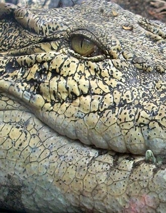
The crocodylian face - once thought to be stony and non-feeling - is sophisticated and covered in sensitive organs, some of which you can see here (they're the tiny dots). This is a Crocodylus porosus. Credit: Darren Naish
Or it could be that facial sensitivity was linked to something like temperature detection? This would presumably be a useful ability as goes the nesting behaviour of these animals. Or what about the possibility that facial sensitivity had a role as goes communication and social life in these dinosaurs? Maybe they rubbed faces during courtship, in family settings, or relayed messages in combat. Crocodylians provide an obvious analogy here, their sensitive facial receptors (the integumentary sense organs – or ISOs – in particular) being involved in the face-stroking and nuzzling that occurs during courtship. Facial bite marks in allosauroids and tyrannosaurids indicate that face-to-face contact was important, possibly having a ritualised combat function (Tanke & Currie 1998, Peterson et al. 2009, Hone & Tanke 2015).
These suggested functions for facial sensitivity are not, of course, mutually exclusive – maybe the enhanced sensitivity of the theropod face allowed them to perform all manner of complex operations and interactions that we primates associate with forelimbs more than faces. How ‘normal’ Neovenator was within the pantheon of theropods as a whole invites further work.
Those contentious extraoral tissues. Does any of this mean anything in particular as goes the life appearance of Neovenator? Those of you familiar with the extinct archosaur research community will be aware of the never-ending debate on dinosaur facial tissues and on whether they had ‘lips’, ‘cheeks’ and other such structures. The latest contribution on this subject was published just a few weeks ago and concerned the new tyrannosaurid species Daspletosaurus horneri (Carr et al. 2017). Therein, it was argued that a rugose skull surface texture – extending all the way to the jaw edges – was linked with scalation and an absence of ‘lips’.
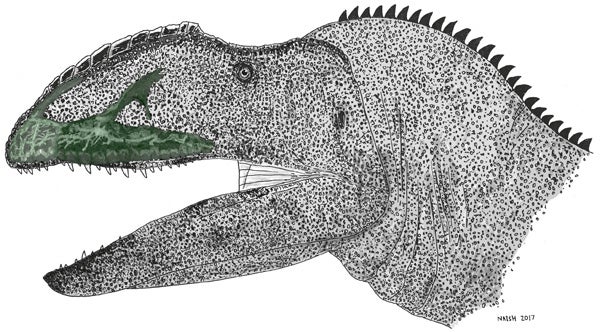
Neovenator life reconstruction with the CT-scanned premaxilla and maxilla shown in situ. Credit: Darren Naish
There’s an aside I have to cover here: Carr et al. (2017) argued that tyrannosaurids were crocodylian-like in having a face covered by “many flat scales”, and they said that the rows of foramina on crocodylian skulls are correlated with “a scaly integument”. This isn’t quite right. The structures that look like scales on a crocodylian face actually represent polygonal cracking of a highly keratinised skin (Milinkovitch et al. 2013). If, ergo, tyrannosaurids shared such a highly keratinised skin with crocodylians, would their very differently shaped skulls generate the same sort of polygonal cracking, and would that cracking – if present – be distributed in the same manner? Large keratinous sheets on the altirostral snout of a big theropod (‘altirostral’ = narrow and tall-sided) might result in a very different look. I’ll leave this matter alone for now.
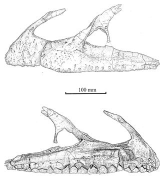
Steve Hutt's excellent drawings of Neovenator's facial bones in left lateral and (below) medial views; note the numerous foramina and sculpted bone texture on the lateral surfaces. These illustrations appeared in Naish et al. (2001) and Brusatte et al. (2008). Credit: Naish et al. 2001
Ultimately, however, our work doesn’t help that much on this issue. The external bone texture and large number of foramina suggests – we think – that a thick external tissue covering was present in Neovenator, and that this covering involved immobile tissue, albeit not rhamphotheca. As we note, even a thick skin is in no way inconsistent with exceptional sensitivity; such is demonstrated by the thick facial epidermis of crocodylians, which is nearly twice as thick as the skin elsewhere on the body.
All that remains for me to say is to thank my co-authors – Chris, Elis, Orestis and Gareth – for all their work on this really fun and exciting project, and to promise that those additional studies on the palaeobiology of Neovenator will one day appear. Not only does this new study enhance our knowledge of extinct dinosaur behaviour and biology, it highlights the fact that even a well described and relatively well-studied species can reveal so much more when subjected to novel analysis.
For previous Tet Zoo articles on big Mesozoic theropods see...
Early abelisaurs and fan-crested and stretch-jawed hadrosaurs
Tyrant dinosaurs were not a Northern Hemisphere speciality: they also colonised Australia!
Concavenator: an incredible allosauroid with a weird sail (or hump)… and proto-feathers?
Refs - -
Brusatte, S. L., Benson, R. B. J. & Hutt, S. 2008. The osteology of Neovenator salerii (Dinosauria: Theropoda) from the Wealden Group (Barremian) of the Isle of Wight. Monograph of the Palaeontographical Society 162, 1-75.
Carr, T. D., Varricchio, D. J., Sedlmayr, J. C., Roberts, E. M. & Moore, J. R. 2017. A new tyrannosaur with evidence with evidence for anagenesis and crocodile-like facial sensory system. Scientific Reports 7, 44942.
Di-Poï, N. & Milinkovitch, M. C. 2013. Crocodylians evolved scattered multi-sensory micro-organs. EvoDevo 4, 1.
Foffa, D., Sassoon, J., Cuff, A. R., Mavrogordato, M. N. & Benton, M. J. 2014. Complex rostral neurovascular system in a giant pliosaur. Naturwissenschaften 101, 453-456.
Hone, D. W. E. & Rauhut, O. W. M. 2010. Feeding behaviour and bone utilization by theropod dinosaurs. Lethaia 43, 232–244.
Hone, D. W. E. & Tanke, D. H. 2015. Pre-and postmortem tyrannosaurid bite marks on the remains of Daspletosaurus (Tyrannosaurinae: Theropoda) from Dinosaur Provincial Park, Alberta, Canada. PeerJ 3, e885.
Ibrahim, N., Sereno, P. C. Dal Sasso, C., Maganuco, S., Fabbri, M., Martill, D. M., Zouhri, S., Myhrvold, N. & Iurino, D. A. 2014. Semiaquatic adaptations in a giant predatory dinosaur. Science 345, 1613-1616.
Leitch, D. B. & Catania, K. C. 2012. Structure, innervation and response properties of integumentary sensory organs in crocodylians. Journal of Experimental Biology 215, 4217–4230.
Milinkovitch, M. C., Manukyan, L., Debry, A., Di-Poï, N., Singh, D., Lambert, D. & Zwicker, M. 2013 Crocodile head scales are not developmental units but emerge from physical cracking. Science 339, 78-81.
Naish, D., Hutt, S. & Martill, D. M. 2001. Saurischian dinosaurs 2: Theropods. In Martill, D. M. & Naish, D. (eds) Dinosaurs of the Isle of Wight. The Palaeontological Association (London), pp. 242-309.
Peterson, J. E., Henderson, M. D., Scherer, R. P. & Vittore, C. P. 2009. Face biting on a juvenile tyrannosaurid and behavioural implications. Palaios 24, 780-784.
Tanke, D. H. & Currie, P. J. 1998. Head-biting behavior in theropod dinosaurs: paleopathological evidence. Gaia 15, 167-184.