This article was published in Scientific American’s former blog network and reflects the views of the author, not necessarily those of Scientific American
A few years ago, as a graduate student in biomedical communications, I completed a unit on embryology as part of an intensive course in human anatomy. Despite the whirlwind nature of the lecture series, which was buried amongst a blur of gross anatomy lessons, hours of dissection, and nerve-wracking lab practicals, it is one of the few subjects on which I’ve actually maintained a modicum of knowledge. Maybe it was the startlingly creepy embryo illustrations that I just couldn’t un-see. Or perhaps it was the singular terminology. Heaven knows I’ll never forget the glorious, fleeting bit of anatomy with which I proudly share my initials, the aorta-gonad-mesonephros.
More likely, though, I suspect it is the sense of pure wonder that accompanies the study of how we come to be. The dizzyingly complex and often mysterious transition from ball of cells to fully formed human, poetically enticing though it may be, is exceptionally difficult to grasp as a student. I believe this is largely due to the simple fact that we cannot see it. Unlike the body of an already developed person, we cannot probe it, scan it, or soberly peel away its layers in the dissection lab. Moreover, even if we could easily photograph it, it is not a static thing. It is a moving, growing evolutionary spectacle. This is where a good illustration becomes a positively invaluable learning tool.
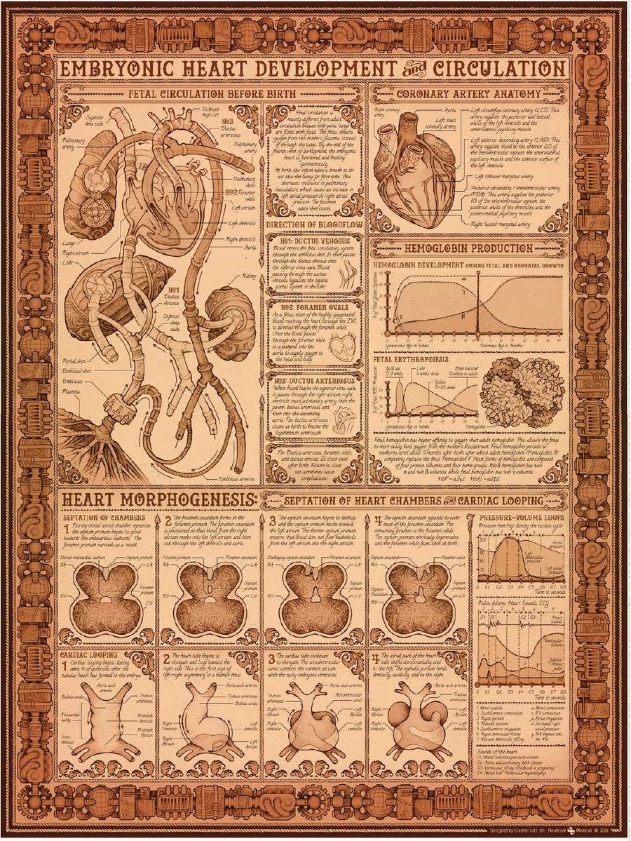
In recent years, Eleanor Lutz has made a name for herself as a science illustrator with a unique ability to visualize, both effectively and accurately, some of the most challenging topics in biology. (You can peruse her website for examples of her work, and in case you missed it, check out the great jellyfish animation she created for Scientific American earlier this year.) To my delight, in her latest work, she takes on one of the most intricate facets of developmental biology: embryonic heart development.
On supporting science journalism
If you're enjoying this article, consider supporting our award-winning journalism by subscribing. By purchasing a subscription you are helping to ensure the future of impactful stories about the discoveries and ideas shaping our world today.

Credit: NERDCORE MEDICAL
In this collaboration with Nerdcore Medical, which you can read more about in her blog post, Eleanor captures the series of chemical processes, structural developments, and precise anatomical convolutions that characterize the formation of the human heart. What’s more, she manages to infuse this complex science with a rich, decorative steam-punk vibe, without compromising visual clarity or educational value. Indeed, these pipe-like blood vessels and florally embellished atria tell the story of heart development as effectively and comprehensively as any textbook illustration I’ve seen. And if I were to go back to that heady and exhausting time in grad school, as I strived to soak up every bit of knowledge about human anatomy and development, you can rest assured I would have this poster on my wall.