This article was published in Scientific American’s former blog network and reflects the views of the author, not necessarily those of Scientific American
In small, shallow splash pools on the coast of the Caribbean island of Curaçao lives a cup-shaped microbe with two neat tricks: it can invert, and it can see.
Although that might not seem so extraordinary from your privileged position in Kingdom Animalia, this multicellular microbe is not an animal. It’s a choanoflagellate, a microbe that feeds on bacteria.
On supporting science journalism
If you're enjoying this article, consider supporting our award-winning journalism by subscribing. By purchasing a subscription you are helping to ensure the future of impactful stories about the discoveries and ideas shaping our world today.
Animals are extremely good at bending sheets of cells, especially during the folding spree of embryonic development. During that time, a single cell divides into many that contort themselves repeatedly to build the many tissues and organs necessary to make a baby. Folding sheets of cells is a foundational animal ability, one vital to their proper construction.
Choanoflagellates are named for their collars of filaments surrounding a beating tail, a combination that produces a rather Sputnik-like profile (as you can see here). They use this equipment to move and eat, as the beating tail can both push the critter and pull in food. In spite their odd appearance, choanoflagellates also happen to be the closest living relatives of animals.
In spite of their kinship to us, prior to the discovery of this new species, dubbed Choanoeca flexa, choanoflagellates had only once before been seen working in teams to bend a sheet of cells. It was no doubt a surprise to the scientists who discovered this little critter that it does so (they published their findings in Science last October, where the discovery made the cover), and they were curious about both how and why it manages the feat.
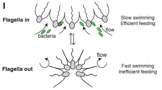
After extensive study, they discovered the following: in C. flexa, the antennae-like collars and flagella line the inside of the cup under most circumstances, when the creature is feeding. During that time, bacteria are pulled by the current of the beating flagella (shown with a wavy line below) toward the bases of the cells, where they can be ingested.

Under certain conditions, they invert, and the now outward-facing tails propel them to a sunnier spot. After a few minutes, the cups relax and revert to their original shape, in which they eat heartily while resting.
The researchers only realized C. flexa could see because they wondered why it inverted. When they tried switching their microscope lights off, the microbes flipped into balls and swam off. This was unexpected. Choanoflagellates were thought to be blind.
So why move, and why see? C. flexa may invert its sheet to escape predators, the scientists speculate, whose shadow gives them away. Or perhaps swimming allows the microbes to gather in bright places where food may be more abundant.
Amazingly, C. flexa not only can "see", it sees using a version of rhodopsin. If that sounds familiar, it is because this is the same protein found in the rods at the back of your eye that help you see in low light. Also just like us, the scientists discovered Choanoeca must eat food that contains either retinal or beta carotene in order to prevent blindness (retinal and beta carotene constitute a rhodopsin building block that neither we nor this choanoflagellate can synthesize).
The similarities don’t end there. The scientists also found that, like other choanoflagellates, C. flexa contains both actin and myosin, the same protein engines that power animal muscles. Even sponges, the houseplants of the animal world, contain these two proteins.
As it turns out, the two C. flexa myosin components are 78% and 63% similar to their human counterparts. When you think about the evolutionary distance involved (over half a billion years), it's a bit mind boggling.
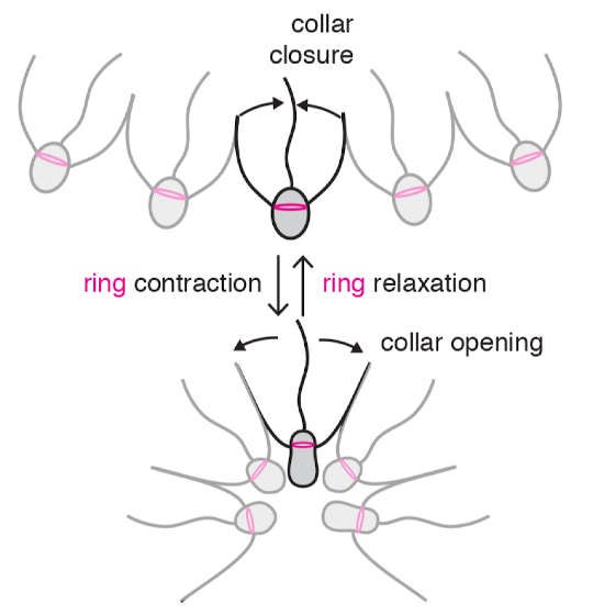
The way in which the actin and myosin are arranged in these cells is also animal-like. Each cell contains actin rings on one end in a configuration analogous to actin rings found in animal tissues called epithelium. They constrict these rings to bend the sheets of cells just as our epithelial cells do. Motion is transmitted through attachment points called cell junctions in animals and, the authors of this new research believe, via touching collar filaments in C. flexa.
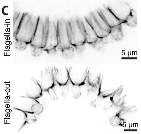
In C. flexa, the actin rings contract and squeeze the top of the cell. This causes the collar fibers to flare from a barrel shape to a cone shape, pressing against one another in such a way that the whole sheet flips. During this time, the cells temporarily change shape from something like bean to something like peanut.

Here's what it looks like under the microscope:

In spite of the similarities to animal tissue folding, such folding in just one or possibly two choanoflagellate species means it probably evolved independently from animals using the commonly inherited apical actin-myosin ring. This circular apparatus is clearly extremely ancient — more than 600 million years old. So C. flexa's ability is still extraordinary, but likely coincidental.
It’s exciting to see a fundamental animal ability in a close relative so different from ourselves. In an embryo, sheet folding is vital to the proper shape and function of an enormously complex animal. In this choanoflagellate, such folding at its very simplest is no less important to the survival of a little microbial oddball.
Reference
Brunet, Thibaut, Ben T. Larson, Tess A. Linden, Mark JA Vermeij, Kent McDonald, and Nicole King. "Light-regulated collective contractility in a multicellular choanoflagellate." Science 366, no. 6463 (2019): 326-334.