This article was published in Scientific American’s former blog network and reflects the views of the author, not necessarily those of Scientific American
Neuroscientists desperately need better tools to develop drugs for treating mental illness and neurodegenerative diseases. Experiments in lab animals more often than not produce drug candidates that ultimately fail in clinical trials. If a laboratory cell culture existed that accurately mimicked the intricate cellular networks that make up the cerebral cortex, it would provide a reality check and help answer the persistent question every drug developer wants to know: if this chemical works in mice that mimic some of the effects of schizophrenia or autism, will it work in humans?
A team from Johns Hopkins Medical School and Nanjing University has just created a microcosm of the cerebral cortex that fits inside a lab dish. The “cortex in a dish” consists of an interwoven mesh of neurons that transmit electrical signals and other cells that damp down this activityThe cultured neurons may expand the toolkit needed to lift neurological and psychiatric drug development out of its present rut.
Scientific American talked with Valina Dawson, co-director of the Institute for Cell Engineering at Johns Hopkins University School of Medicine, about a paper published April 6 in Science Translational Medicine on which she was the senior author.
On supporting science journalism
If you're enjoying this article, consider supporting our award-winning journalism by subscribing. By purchasing a subscription you are helping to ensure the future of impactful stories about the discoveries and ideas shaping our world today.
Scientific American: Describe what a 'cortex in a dish' is?
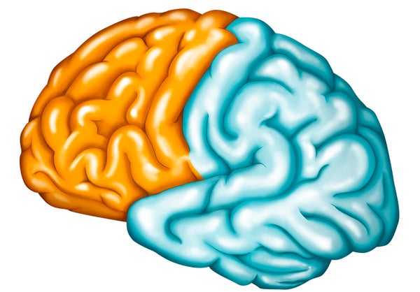
Valina Dawson: The brain is divided into different structures and regions. The cerebral cortex is the largest part of the brain and manages higher brain functions such as thought and actions, language, sensory processing such as hearing and vision. One way to study how the neurons in the cortex function is to grow them in culture, “in a dish.”
SA: Why have researchers wanted to create this laboratory model of the cortex?
VD: Having human neurons in culture allows experimental investigations into signaling events at the chemical, protein and genetic levels that underlie normal and diseased actions in a manner that would not be possible or ethical in an intact human brain. Understanding how the neurons in the cortex work and what goes wrong when disease occurs will provide, we hope, new therapeutic opportunities to treat patients who suffer from brain injury and disease.
SA: Why has it been so difficult to build this and how did you overcome hurdles along the way?
VD: The ability to create human neurons from stem cells is relatively new. Established protocols to make different types of neurons from stem cells are limited to a few types of neurons out of the hundreds that exist in the human brain. Most of our understanding about the carefully controlled and elegantly choreographed events in the development of the cortex are from less complex organisms. Thus some of the key elements necessary to generate a protocol to produce the complex network of neuronal populations had to be guessed and identified by trial and error.
SA:. What will scientists be able to do with your system?
VD: We hope they will use this system to understand important mechanisms in cortical function and communication. We hope these cultures will also be useful in understanding how to provide neuroprotection against stroke and trauma and to investigate what goes wrong in diseases such as schizophrenia, autism and epilepsy.
SA: Didn’t you show in the paper just published how this might work?
Yes, in a model of stroke. Stroke is a common cause of death, disability and loss of quality of life world-wide but unfortunately there are few treatments to reduce the brain injury suffered. During a stroke there is a loss of blood flow to brain tissue. This can be mimicked in a culture dish by removing oxygen and glucose or by activating with the chemical NMDA a specific protein on the surface of neurons, an excitatory receptor. We have used our cortical culture system to study the cellular signaling events that occur and lead to neuronal cell death. Previously we found in rodent systems, that a member of a biochemical pathway, poly(ADP-Ribose) polymerase-1 (PARP-1) is pathologically activated and serves as a switch, directing the cell away from DNA repair towards cell death.
Preventing PARP-1 activation protects neurons from ischemic cell death. For the first time we were able to determine using our cortical culture if human neurons respond in a similar manner, and they do! Inhibitors of PARP have been developed for the treatment of patients with certain types of cancer and some of these clinically useful drugs cross the blood brain barrier. Finding agents that can gain access to the brain has been a major hurdle in developing good treatments for neurologic disease and injury. Our studies raise the potential that these drugs could also be useful in the treatment of stroke.
SA: What are the next steps?
VD: Besidesusing these cultures to study how cortical neurons protect themselves from injury, we are also using these cultures to probe the molecular signaling events that underlie a form of autism with the hopes of finding a way to intervene. Additionally, other brain regions connect to the cortex, providing important information or receiving instructions from cortical regions. In the future, cultures could be established so that these connections could be studied at the cellular and sub-cellular level. One could also envision using sophisticated bioengineered scaffolds to permit the normal layering of the cortex so that interconnectivity between layers could be studied. In the science fiction future, perhaps cortical plugs would be developed that could be implanted into patients with stroke or trauma to replace the brain material that was damaged and lost.